18 Epithelial Tissue
Characteristics of Epithelial Tissue
The human body consists of four types of tissue: epithelial, connective, muscular, and nervous. Epithelial tissue covers the body, lines all cavities, and composes the glands.
Learning Objectives
Describe the primary functions and characteristics of epithelial tissue
Key Takeaways
Key Points
- Epithelial tissue is composed of cells laid together in sheets with the cells tightly connected to one another. Epithelial layers are avascular, but innervated.
- Epithelial cells have two surfaces that differ in both structure and function.
- Glands, such as exocrine and endocrine, are composed of epithelial tissue and classified based on how their secretions are released.
Key Terms
- epithelium: A membranous tissue composed of one or more layers of cells that form the covering of most internal and external surfaces of the body and its organs.
- avascular: Lacking blood vessels.
- vascular: Containing blood vessels.
Functions of the Epithelium
Epithelia tissue forms boundaries between different environments, and nearly all substances must pass through the epithelium. In its role as an interface tissue, epithelium accomplishes many functions, including:
- Protection for the underlying tissues from radiation, desiccation, toxins, and physical trauma.
- Absorption of substances in the digestive tract lining with distinct modifications.
- Regulation and excretion of chemicals between the underlying tissues and the body cavity.
- The secretion of hormones into the blood vascular system. The secretion of sweat, mucus, enzymes, and other products that are delivered by ducts come from the glandular epithelium.
- The detection of sensation.
Characteristics of Epithelial Layers
Epithelial tissue is composed of cells laid out in sheets with strong cell-to-cell attachments. These protein connections hold the cells together to form a tightly connected layer that is avascular but innervated in nature.
The epithelial cells are nourished by substances diffusing from blood vessels in the underlying connective tissue. One side of the epithelial cell is oriented towards the surface of the tissue, body cavity, or external environment and the other surface is joined to a basement membrane. The basement layer is non-cellular in nature and helps to cement the epithelial tissue to the underlying structures.
Types of Epithelial Tissue
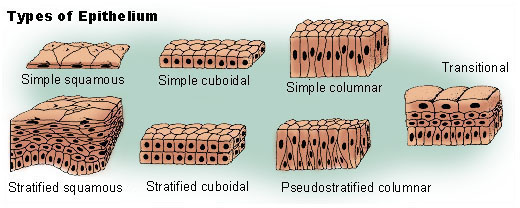
Epithelial tissues are identified by both the number of layers and the shape of the cells in the upper layers. There are eight basic types of epithelium: six of them are identified based on both the number of cells and their shape; two of them are named by the type of cell (squamous) found in them. Epithelial tissue is classified based on the number of cells, the shape of those cells, and the types of those cells.
| Epithelial Tissue Cells | ||
|---|---|---|
| Cells | Locations | Function |
| Simple squamous epithelium
|
Air sacs of the lungs and the lining of the heart, blood vessels and lymphatic vessels | Allows materials to pass through by diffusion and filtration, and secretes lubricating substances |
| Simple cuboidal epithelium
|
In ducts and secretory portions of small glands and in kidney tubules | Secretes and absorbs |
| Simple columnar epithelium
|
Ciliated tissues including the bronchi, uterine tubes, and uterus; smooth (nonciliated tissues) are in the digestive tract bladder | Absorbs; it also secretes mucous and enzymes. |
| Pseudostratified columnar epithelium
|
Ciliated tissue lines the trachea and much of the upper respiratory tract | Secrete mucous; ciliated tissue moves mucous |
| Stratified squamous epithelium
|
Lines the esophagus, mouth, and vagina | Protects against abrasion |
| Stratified cuboidal epithelium
|
Sweat glands, salivary glands, and mammary glands | Protective tissue |
| Stratified columnar epithelium
|
The male urethra and the ducts of some glands. | Secretes and protects |
| Transitional epithelium
|
Lines the bladder, urethra and ureters | Allows the urinary organs to expand and stretch |
Types of Epithelial Tissue
Epithelial tissue is classified by cell shape and the number of cell layers.
Learning Objectives
Classify epithelial tissue by cell shape and layers
Key Takeaways
Key Points
- There are three principal cell shapes associated with epithelial cells: squamous epithelium, cuboidal epithelium, and columnar epithelium.
- There are three ways of describing the layering of epithelium: simple, stratified, and pseudostratified.
- Pseudostratified epithelium possesses fine hair-like extensions called cilia and unicellular glands called goblet cells that secrete mucus. This epithelium is described as ciliated pseudostratified epithelium.
- Stratified epithelium differs from simple epithelium in that it is multilayered. It is therefore found where body linings have to withstand mechanical or chemical insult.
- In keratinized epithelia, the most apical layers (exterior) of cells are dead and and contain a tough, resistant protein called keratin. An example of this is found in mammalian skin that makes the epithelium waterproof.
- Transitional epithelia are found in tissues such as the urinary bladder where there is a change in the shape of the cell due to stretching.
Key Terms
- simple columnar: A columnar epithelium that is uni-layered.
- pseudostratified epithelium: A type of epithelium that, though comprising only a single layer of cells, has its cell nuclei positioned in a manner suggestive of stratified epithelia.
- squamous: Flattened and scale-like.
- cuboidal: Resembling a cube.
- Keratinized: To produce or become like keratin.
- columnar: Having the shape of a column.
Most epithelial tissue is described with two names. The first name describes the number of cell layers present and the second describes the shape of the cells. For example, simple squamous epithelial tissue describes a single layer of cells that are flat and scale-like in shape.

Epithelial Tissue: There are three principal classifications associated with epithelial cells. Squamous epithelium has cells that are wider than they are tall. Cuboidal epithelium has cells whose height and width are approximately the same. Columnar epithelium has cells taller than they are wide.
Simple Epithelia
Simple epithelium consists of a single layer of cells. They are typically where absorption, secretion and filtration occur. The thinness of the epithelial barrier facilitates these processes.
Simple epithelial tissues are generally classified by the shape of their cells. The four major classes of simple epithelium are: 1) simple squamous; 2) simple cuboidal; 3) simple columnar; and 4) pseudostratified.
Simple Squamous
Simple squamous epithelium cells are flat in shape and arranged in a single layer. This single layer is thin enough to form a membrane that compounds can move through via passive diffusion. This epithelial type is found in the walls of capillaries, linings of the pericardium, and the linings of the alveoli of the lungs.
Simple Cuboidal
Simple cuboidal epithelium consists of a single layer cells that are as tall as they are wide. The important functions of the simple cuboidal epithelium are secretion and absorption. This epithelial type is found in the small collecting ducts of the kidneys, pancreas, and salivary glands.
Simple Columnar
Simple columnar epithelium is a single row of tall, closely packed cells, aligned in a row. These cells are found in areas with high secretory function (such as the wall of the stomach), or absorptive areas (as in small intestine ). They possess cellular extensions (e.g., microvilli in the small intestine, or the cilia found almost exclusively in the female reproductive tract).
Pseudostratified
These are simple columnar epithelial cells whose nuclei appear at different heights, giving the misleading (hence pseudo) impression that the epithelium is stratified when the cells are viewed in cross section.
Pseudostratified epithelium can also possess fine hair-like extensions of their apical (luminal) membrane called cilia. In this case, the epithelium is described as ciliated pseudostratified epithelium. Ciliated epithelium is found in the airways (nose, bronchi), but is also found in the uterus and fallopian tubes of females, where the cilia propel the ovum to the uterus.
Stratified Epithelium
Stratified epithelium differs from simple epithelium by being multilayered. It is therefore found where body linings have to withstand mechanical or chemical insults.
Stratified epithelia are more durable and protection is one their major functions. Since stratified epithelium consists of two or more layers, the basal cells divide and push towards the apex, and in the process flatten the apical cells.
Stratified epithelia can be columnar, cuboidal, or squamous type. However, it can also have the following specializations:
Keratinized Epithelia
In keratinized epithelia, the most apical layers (exterior) of cells are dead and lose their nucleus and cytoplasm. They contain a tough, resistant protein called keratin. This specialization makes the epithelium waterproof, and it is abundant in mammalian skin. The lining of the esophagus is an example of a non-keratinized or moist stratified epithelium.
Transitional Epithelia
Transitional epithelia are found in tissues that stretch and it can appear to be stratified cuboidal when the tissue is not stretched, or stratified squamous when the organ is distended and the tissue stretches. It is sometimes called the urothelium since it is almost exclusively found in the bladder, ureters, and urethra.


