25 Accessory Structures of the Skin
Sweat (Sudoriferous) Glands
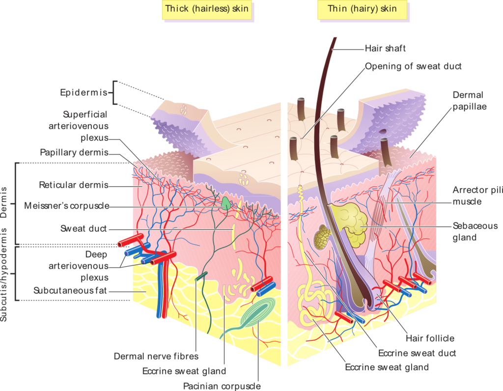
Sweat glands, also known as sudoriferous glands, are distributed over most of the body surface.
Learning Objective
Classify eccrine and apocrine sweat glands
Key Takeaways
Key Points
- Sweat glands are located deep within the skin and primarily regulate temperature.
- The two main types of sweat glands are eccrine sweat glands and apocrine sweat glands.
- Eccrine sweat glands are smaller sweat glands. They are coiled tubular glands that discharge their secretions directly onto the surface of the skin.
- Apocrine sweat glands are coiled tubular glands that discharge in the canals of hair follicles. The sweat produced may be acted upon by bacteria, causing a noticeable odor.
Key Terms
- eccrine gland: The major sweat glands of the human body, found in virtually all skin, produce a clear, odorless substance, consisting primarily of water and NaCl.
- apocrine sweat gland: The type of sweat gland that is least responsible for thermoregulation and most responsible for body odor.
Sweat glands, also called sudoriferous glands, are simple tubular glands found almost everywhere on our body. Each sweat gland is made up of two portions:
- A secretory section
- An excretory duct
The secretory portion is found in the dermis, the middle layer of the skin. Sometimes it’s also found in the hypodermis, the deepest layer of our skin.
The secretory portion of a sweat gland is a twisted and coiled tube that has an opening at its very top. It is in the coiled secretory portion of the sweat gland where the sweat is actually produced. The excretory duct moves from the secretion portion, through the dermis, and into the topmost layer of the skin, the epidermis, where it opens up at the surface of our skin.
Eccrine Glands

The most numerous types of sweat glands in our skin, found almost everywhere on the body, are called eccrine glands. These are the true sweat glands in the sense of helping to regulate body temperature. In other words, sweating causes the loss of body heat and thus cools us down on a hot day or when performing strenuous exercise. This is because as the water in sweat evaporates, it takes body heat with it.
Apocrine Glands
The other kind of sweat glands are known as apocrine glands. The apocrine glands are found in places like the armpits, scrotum, anus, and labia majora. They are typically larger than eccrine glands and their ducts tend to open into hair follicles instead of hairless areas of skin.
These glands, unlike the eccrine glands, serve virtually no role in the regulation of body temperature. These are also the glands largely responsible for body smells, as their excretions are converted by skin bacteria into various chemicals we associated with body odor.
Unlike eccrine glands, the exact function of apocrine glands is unknown and debated. We do know they are activated during times of stress, pain, and sexual foreplay but for what reasons is yet to be made clear.
Sebaceous (Oil) Glands
Sebaceous glands are found in most of the skin (except the palms of the hands and the soles of the feet).
Learning Objective
Describe the location and function of sebaeous glands
Key Takeaways
Key Points
- Sebaceous glands are located throughout the skin except in the palms of the hands and soles of the feet.
- Sebum is an oily substance composed of fat (lipids) and the debris of dead fat-producing cells.
- Sebaceous glands are classified as holocrine glands.
Key Terms
- sebum: A thick oily substance, secreted by the sebaceous glands of the skin, that consists of fat and cellular debris.
- holocrine gland: The sebaceous gland is an example of a holocrine gland because its product of secretion (sebum) is released with remnants of dead cells.
Sebaceous glands are the oil secreting glands of your body. This is why they are also called the oil glands. They are a type of holocrine simple saccular (alveolar) gland. Their function is to secrete a substance called sebum, a mixture of fatty substances, entire sebum-producing cells, and epithelial cell debris. The sebaceous glands are located in the dermis, the middle layer of the skin, and they develop from the epithelial cells of the hair follicle itself (the external root sheath of the hair follicle).
Sebaceous gland ducts thus usually open up into the upper part of a hair follicle, called the infundibulum. The infundibulum is part of the pilosebaceous canal, the one responsible for discharging sebum and one that is composed of the infundibulum and the short duct of the sebaceous gland itself. However, some sebaceous gland ducts open directly onto our skin surface such as at the corner of the mouth and the glans penis. Regardless, the secretion of sebum out of the gland is helped along by the contraction of the arrector pili muscle.
While the sebaceous glands are present just about all over the skin, they are notably absent on the palms of the hands and the soles of the feet. The sebum being excreted by your body today began production around 8 days ago.
The Function of Sebum

The sebum produced by these glands plays numerous important roles:
- Sebum is a lubricant and inasmuch it helps to moisturize the skin. It does so by preventing the excess evaporation of water from the skin.
- Sebum serves to keep us healthy by keeping in check the growth of certain bacteria on our skin. That’s because sebum contains chemicals that kill bacteria. This helps ensure bacteria don’t invade into deeper layer of our skin.
- It helps to condition the hair. Meaning, it ensures our hair doesn’t become too dry and brittle.
Sebaceous glands are involved in numerous conditions. During puberty, various hormones cause them to produce a lot of sebum and this therefore contributes to oily skin. If a duct of a sebaceous gland is clogged with sebum, a whitehead results. If this material is allowed to dry and oxidize, it will become darker, forming a blackhead. If a sebaceous gland becomes infected, moderate and severe forms of acne are the result.
The glands lining the ear canal that produce earwax (cerumen) are called ceruminous glands. They are modified sebaceous glands.
Nails
Fingernails are made of keratin and they perform two major functions: protection and sensation.
Learning Objective
Describe the structure of fingernails
Key Takeaways
Key Points
- The nail bed contains the blood vessels, nerves, and melanocytes or melanin-producing cells. As the nail is produced by the root, it streams down along the nail bed, which adds material to the undersurface of the nail and makes it thicker.
- The nail plate is the actual fingernail, composed of translucent keratin. The pink appearance of the nail comes from the blood vessels underneath the nail.
- The eponychium, or cuticle, is situated between the skin of the finger and the nail plate. It fuses these structures together and provides a waterproof barrier.
- Deformity or disease of the nails is referred to as onychosis. There are many diseases that can occur with the fingernails and toenails. The most common of these diseases are ingrown nails and fungal infections.
- Ingrown nails, also known as onychocryptosis, can affect either the fingers or the toes. In this condition, the nail cuts into one or both sides of the nail bed, resulting in inflammation and possibly infection.
Key Term
- keratin: A protein that makes up hair and nails.
Function of the Fingernail
The fingernail is an important structure made of keratin. The fingernail generally serves two purposes: it acts as a protective plate and enhances sensation of the fingertip. Nails can also help grasp small things.
The protection function of the fingernail is commonly known, but the sensation function is equally important. The fingertip has many nerve endings in it that allow it to receive volumes of information about the objects we touch. The nail acts as a counterforce to the fingertip, providing even more sensory input when an object is touched.
Nails grow from the nail bed continuously but they slow down their growth rate with age, poor nutrition, or poor circulation.
Anatomy of the Fingernail
The structure of the fingernail is divided into six specific parts:
- root
- nail bed
- nail plate
- eponychium (cuticle)
- perionychium
- hyponychium
Root and Nail Sinus
The nail sinus (sinus unguis) is where the nail root is—at the base of the nail underneath the skin. It originates from the actively growing tissue below, the matrix. The root of the fingernail is also known as the germinal matrix.
This portion of the nail is actually beneath the skin, behind the fingernail, and extends several millimeters into the finger. The fingernail root produces most of the volume of the nail and the nail bed. This portion of the nail does not have any melanocytes, or melanin-producing cells. The edge of the germinal matrix is seen as a white, crescent shaped structure called the lunula.
Nail Bed
The nail bed is a part of the nail matrix called the sterile matrix. It extends from the edge of the germinal matrix, or lunula, to the hyponychium. The nail bed contains the blood vessels, nerves, and melanocytes, or melanin-producing cells. As the nail is produced by the root, it streams down along the nail bed, which adds material to the undersurface of the nail and makes it thicker.
Nail Plate
The nail plate is the actual fingernail, composed of translucent keratin. The pink appearance of the nail comes from the blood vessels underneath the nail. The underneath surface of the nail plate has grooves along the length of the nail that help anchor it to the nail bed. The free margin or distal edge is the anterior margin of the nail plate corresponding to the abrasive or cutting edge of the nail.
Eponychium
The eponychium, or cuticle, is situated between the skin of the finger; the nail plate fuses these structures together and provides a waterproof barrier.
Perionychium
The perioncyhium is the skin that overlies the nail plate on its sides; it is also known as the paronychial edge. The perionychium is the site of hangnails, ingrown nails, and an infection of the skin called paronychia.
Hyponychium
The hyponychium is the area between the nail plate and the fingertip. It is the junction between the free edge of the nail and the skin of the fingertip, also providing a waterproof barrier.
Hair
Hair growth occurs from the hair follicle.
Learning Objective
Describe the characteristics of body hair
Key Takeaways
Key Points
- Hair primarily serves for protection, warmth, and sensation.
- Human hair is made of keratin.
- The structure of the hair follicle includes the papilla, matrix, root, and bulb.
- The different types of hair on the human body include lanugo, vellus hair, and terminal hair.
- Attached to the follicle is a tiny bundle of muscle fiber called the arrector pili.
Key Terms
- hair follicle: The structure that produces hair.
- papilla: A structure that provides nutrients that help our hair grow.
Hair is present on almost the entire surface of our body, excluding certain regions such as the palms of our hands, soles of our feet, and some genital areas. In certain places, hair is so small that it is virtually invisible to the naked eye and in other places it is quite obvious, like on our head and in our armpits.
Structure
A hair can be divided into two main parts lengthwise:
- The root is part of the hair enclosed by the hair follicle, which is itself a tube-like involution of the skin.
- The shaft is the part of the hair projecting from the surface of our skin. A round shaft results in straight and coarse hair. An oval hair shaft is responsible for wavy hair. A flat shaft causes curly hair.
Each hair can also be broken into three main parts. They are, from the most superficial to the deepest:
- Cuticle, the outermost portion. This layer is composed of scale-like cells that seem to overlap in a shingle-like manner. Such an arrangement helps prevent hair from matting.
- Cortex, the middle portion. This portion is made up of layers of elongated and flattened cells.
- Medulla, the inner (central) portion. This is made up of large cells with many sides and air spaces.
There are three types of hairs:
- Lanugo, found on the fetus and for a bit after birth.
- Vellus, fine body hair (peach-fuzz hair).
- Terminal hair, the coarse hair.
Growth and Composition
In the deepest portion of the each hair follicle lies the hair bulb. It is supplied with nutrients via blood by a structure called the hair papilla. The hair bulb has a growth zone called the matrix. Here, stratum basale epithelial cells divide via mitosis to form the hair.
As new cells form, they push older cells up to the surface. These older cells die and become keratinized in the process. This means that most hair is made up of protein (keratin).
The hair bulb also contains cells called melanocytes that produce various kinds of melanin pigments. Their varying combinations are what help to produce the different natural hair colors people have.
Hair grows in three stages:
- Anagen phase, the fast growing phase.
- Catagen phase, the involution phase.
- Telogen phase, the rest phase.
Purpose
Depending on the kind of hair and location, hair can have one of several purposes:
- Expression. Hair on our eyebrows may have stuck around on our body to help gauge a person’s emotions or intent.
- Protection. Eyelashes help protect our eyes and nose hairs keep things out of our respiratory system. Hair on the head helps add a small cushion against bumps.
- Sensation. Hair helps us sense light touches.
- Warmth and defense. Attached to a hair follicle is a bundle of muscle fibers. They help comprise the arrector pili muscle that causes the hairs on our body to stand on their ends when we’re cold or we’re scared. The standing of the hair on end also dimples the skin and thus produces goosebumps. In non-human mammals this action may help add an insulating layer of air between the hair (called fur in non-human mammals) and skin, or it may be used as a way to make a scared animal look larger to its enemy. Neither function is all that relevant or useful in humans, although some warmth is provided by scalp hair.