24 Cell Junctions
Tight Junctions
Tight junctions serve as selectively permeable seals in our body’s internal and external surfaces.
Learning Objective
Describe the characteristics of tight junctions
Key Takeaways
Key Points
- Tight junctions are the closely associated areas of two cells whose membranes join together to form a virtually impermeable barrier to fluid.
- Tight junctions perform vital functions—such as holding cells together—and form protective and functional barriers.
- Tight junctions are composed of a branching network of sealing strands with each strand acting independently from the others.
- The major types of proteins in junctions are the claudins and the occludins.
- Each strand is formed from a row of transmembrane proteins embedded in both plasma membranes, with extracellular domains joining one another directly.
Key Terms
- blood-brain barrier: A structure in the central nervous system (CNS) that keeps various substances found in the bloodstream out of the brain while allowing in the substances essential to metabolic function, e.g., oxygen.
- Claudins: Proteins that form the backbone of the tight junction strands.
- cell adhesion molecule: Molecules that help cells stick to each other and to their surroundings. The proteins located on the cell surface bind with other cells or with the extracellular matrix (ECM).
- cytoskeleton: A cellular structure like a skeleton, contained within the cytoplasm.
- epithelia: The covering of internal and external body surfaces, where tight junctions are found.
- zonula occludens: Another name for tight junctions.

Imagine a largely waterproof zipper connecting the sides of two different jackets. That zipper is like a tight junction (TJ), also called an occluding junction. A TJ creates a small zone that occludes the extracellular space (the space between cells).
This is why tight junctions are also called zonula occludens. The word zonula comes from words that mean small zone or encircling belt, while occludens comes from the Latin word occludere, which means to close up.
Location and Function
Tight junctions are virtually (but also partly selectively) impermeable seals that encircle cells and bind them together into leakproof sheets. In other words, the plasma membranes of adjacent cells essentially fuse together tightly in order to limit the leakage of various substances between the two cells.
What can and cannot go through all depends on the substance’s size, charge, as well as the location and precise composition of the tight junctions in the part of the body in question.
Tight junctions are located within our body’s epithelia. Epithelia is the plural of epithelium. Epithelium is a word that refers to the covering of the body’s internal and external surfaces. This includes organs (such as skin), blood vessels, and cavities.

Thus, these tight junctions serve various functions, depending on what epithelium is in question. In the skin, they keep us somewhat watertight and help keep allergens out of our body. In the digestive system, they help prevent the leakage of digestive enzymes into our bloodstream.
Tight junctions also serve as a structural support mechanism that help keep the epithelium together.
Composition
A tight junction—a kind of symmetrical cell junction—is composed of numerous important proteins that are either directly involved in its composition or intimately involved with connecting the tight junction to and between the cells in one way or another. These proteins include:
- Occludins, which maintain the barrier between adjacent cells.
- Claudins, which form the backbone of tight junction strands.
- Junctional adhesion molecules (JAMs) are immunoglobulin (antibody) proteins that help seal the intercellular space between two cells.
- Zonula occludens (ZO) are proteins that help link the tight junction to each cell’s internal skeleton (cytoskeleton).
The occludins and claudins are the major components of tight junction strands. When fully formed, a tight junction is not one, long, continuous seal. Instead, it looks like a series of local seals joined together in a maze-like fashion.

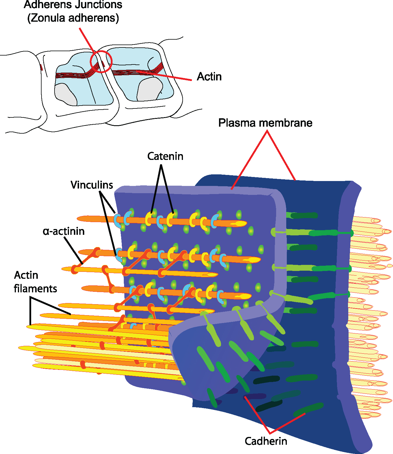
Adherens Junctions
Adherens junctions provide strong mechanical attachments between adjacent cells through the linkage of cytoplasmic face with cytoskeleton.
Learning Objective
Describe the characteristics of adherens junctions
Key Takeaways
Key Points
- Adherens junctions are involved in a number of critical functions, including providing additional structural support. For example, they hold cardiac muscle cells tightly together as the heart expands and contracts.
- Adherens junctions are built primarily from cadherins, whose extracellular segments bind to each other and whose intracellular segments bind to catenins. Catenins are connected to actin filaments.
Key Terms
- cadherin: Any of a class of transmembrane proteins important in maintaining tissue structure.
- adherens junctions: Protein complexes that occur at cell–cell junctions in epithelial tissues; they are usually more basal than tight junctions.
- catenin: Any of a class of proteins that have a role in cell adhesion.
Adherens junctions are also referred to as zonula adherens, intermediate junction, or as belt desmosomes. Zonula means small zone or belt-like, and adherens refers to adhesion (sticking together). As a result, the zonula adherens often runs like a belt around the entire cell in a continuous fashion, and it acts as an adhesion belt.
Location and Function
This type of cell junction is located right below tight junctions and provides a strong bond between the sides of adjacent epithelial cell membranes. While other junctions, like tight junctions, provide some support for and fusion of adjacent cells, their resistance to mechanical stress is relatively small compared to the much stronger adherens junctions.
Structure and Composition
The zonula adherens is composed of several different proteins:
- The actin microfilaments of the cytoskeleton (the internal skeleton of the cell).
- Anchor proteins, found inside each cell. These are called alpha-catenin, beta-catenin, gamma-catenin (aka plakoglobin), vinculin, and alpha-actinin. They link the actin microfilaments to the cadherins.
- Cadherins, namely E-cadherin. These are transmembrane adhesion proteins, whose main portions are located in the extracellular space.
The extracellular part of one cell’s cadherin binds to the extracellular part of the adjacent cell’s cadherin in the space between the two cells. Each cell’s cadherin molecule also contains a tail that inserts itself inside its respective cell.
This intracellular (within the cell) tail then links up to catenin proteins to form the cadherin–catenin complex. This complex binds to vinculin and alpha-actinin; these two proteins are what link the cadherin–catenin complex to the cell’s internal skeletal framework (the actin microfilaments).
The extracellular portions of the cadherin molecules of adjacent cells are bonded together by calcium ions (or another protein in some cases). This means that the functional as well as morphological integrity of the adherens junctions are calcium dependent. If you were to remove calcium from the equation, this type of cell junction would disintegrate as a result.
Gap Junctions
A gap junction is a specialized cell junction that directly connects the cytoplasm of two cells.
Learning Objective
Describe the characteristics of gap junctions
Key Takeaways
Key Points
- Gap junctions allow various molecules and ions to pass freely between cells.
- A gap junction channel is composed of two connexons, also known as hemichannels that line up across the intercellular space.
- Most gap junction hemichannels are composed of a complex of six connexin proteins, each characterized by four transmembrane domains. Six connexin sub-units assemble to create one connexon, or hemichannel.
- Channel composition influences the function of the gap junction.
- Gap junctions allow for electrical communication between cells, and also allow the passage of small second messengers.
- Gap junctions are expressed in virtually all tissues and cells, but most notably in cell types that are involved in direct electrical communication, such as neurons and cardiac muscle.
Key Terms
- cytoplasm: The contents of a cell except for the nucleus. It includes cytosol, organelles, vesicles, and the cytoskeleton.
- connexin
- connexon
- nexus: An alternative name for a gap junction.
Gap junctions are also called communicating junctions, macula communicans, or nexuses. These are connections that allow for the direct passage of molecules between two cells.
Gap junctions consist of a number of transmembrane channels called pores that are found in a closely packed arrangement. The number of gap junctions shared between two cells can vary as well.
Structure
Each gap junction channel is made up of two half channels (hemichannels), one in each cell’s membrane. These half channels join together, bridge the extracellular space in the process, and form the entire channel that spans both cell membranes.
Each of these half channels is called a connexon. Each connexon is made up of six symmetrical integral membrane protein units called connexins. This means each channel is made up of 12 circularly arranged protein units.
Function

The molecules that may cross this channel include the likes of ions, regulatory proteins, and metabolites (products of metabolism). Examples of this includes calcium ions and cAMP (cyclic adenosine monophosphate).
Depending on the type of gap junction in question, molecules can pass evenly in both directions, or asymmetrically, so in some gap junctions the molecules will move in one direction faster than in the other direction.
The channels in a gap junction aren’t always open. They fluctuate between being open and closed. The ability of the channel to open or close is made possible in part to calcium ions, which induce a reversible conformational change in the connexin molecules, which leads to the closure of a channel at its extracellular surface. The cytoplasmic end of each connexon can also be closed, if necessary.
Location
Gap junctions are found in many places throughout the body. This includes epithelia, which are the coverings of body surfaces, as well as nerves, cardiac (heart) muscle, and smooth muscle (such as that of the intestines).
Their primary role is to coordinate the activity of adjacent cells. For instance, when heart cells need to beat in unison, gap junctions allow for the transmission of electrical signals between the cells.

