191 Changes to the Mother’s Body During Pregnancy
Anatomical Changes
Women undergo many physical changes during pregnancy due to hormonal fluctuations and the need to accommodate a growing fetus.
Learning Objectives
Describe the anatomic changes to a pregnant woman that occur over the course of the pregnancy
Key Takeaways
Key Points
- As the fetus grows and develops, several anatomical changes must occur to the female body to accommodate the growing fetus, including placental development, weight gain, abdominal extension, breast enlargement, glandular development, and posture changes.
- During the second trimester, morning sickness subsides, the uterus expands up to 20 times its normal size, breasts enlarge, and movements of the fetus may be felt.
- During the third trimester the fetus grows most rapidly and final weight gain occurs. The abdomen drops and fetal movement can become quite strong. The woman feels ready to give birth.
Key Terms
- implantation: The attachment of the fertilized ovum to the uterine wall.
- decidua: A mucous membrane that lines the uterus that is shed during menstruation and modified during pregnancy.
- pelvic cavity: A body cavity that is bounded by the bones of the pelvis. Its oblique roof is the pelvic inlet (the superior opening of the pelvis). Its lower boundary is the pelvic floor.
Physical Changes During Pregnancy
Pregnancy begins when the developing embryo implants in the endometrial lining of a woman’s uterus. Most pregnant women do not have any specific signs or symptoms after implantation, although it is not uncommon to experience minimal bleeding.
After implantation, the uterine endometrium is called the decidua. The placenta, which is partly formed from the decidua and partly from outer layers of the embryo, connects the developing embryo to the uterine wall to allow nutrient uptake, waste elimination, and gas exchange via the mother’s blood supply.
The umbilical cord connects the embryo or fetus to the placenta. The developing embryo undergoes tremendous growth and changes during the process of fetal development.
Most pregnant women experience a number of symptoms that can signify pregnancy. The symptoms include nausea and vomiting, excessive tiredness and fatigue, cravings for certain foods that are not normally sought out, and frequent urination, particularly during the night.
A number of early medical signs are associated with pregnancy. These signs typically appear, if at all, within the first few weeks after conception. Not all of these signs are universally present, nor are all of them diagnostic by themselves; taken together, however, they may make a presumptive diagnosis of pregnancy.
These signs include:
- The presence of human chorionic gonadotropin (hCG) in the blood and urine.
- Missed menstrual period.
- Implantation bleeding (occurs at implantation of the embryo in the uterus during the third or fourth week after last menstrual period).
- Increased basal body temperature sustained for over 2 weeks after ovulation.
- Chadwick’s sign (darkening of the cervix, vagina, and vulva).
- Goodell’s sign (softening of the vaginal portion of the cervix).
- Hegar’s sign (softening of the uterus isthmus).
- Pigmentation of the linea alba (called linea nigra), which is darkening of the skin in a midline of the abdomen. This darkening is caused by hyperpigmentation resulting from hormonal changes, usually appearing around the middle of pregnancy.
Breast tenderness is common during the first trimester. Shortly after conception, the nipples and areolas begin to darken due to a temporary increase in hormones. This process continues throughout the pregnancy.
Despite all the signs, some women may not realize they are pregnant until they are far along in pregnancy. In some cases, a few have not been aware of their pregnancy until they begin labor. This can be caused by many factors, including irregular periods (quite common in teenagers), certain medications (not related to conceiving children), and obese women who disregard the pregnancy-related weight gain. Others may be in denial of their situation.
The First Trimester
The first 12 weeks of pregnancy are known as the first trimester. During this trimester, fetal development can be divided into different stages.
At the beginning of the fetal stage, the risk of miscarriage decreases sharply. All major structures, including the head, brain, hands, feet, and other organs have been formed by the fetal stage. Once pregnancy moves into the second trimester, the risks of miscarriage and birth defects drops drastically.
The Second Trimester
Weeks 13 to 28 of the pregnancy are called the second trimester. Most women feel more energized in this period. They put on weight as the symptoms of morning sickness subside and eventually fade away.
By the end of the second trimester, the expanding uterus has created a visible baby bump. Although the breasts have been developing internally since the beginning of the pregnancy, most of the visible changes appear after this point.
The uterus can expand up to 20 times its normal size during pregnancy. Although the fetus begins to move and takes a recognizable human shape during the first trimester, it is not until the second trimester that movement of the fetus, often referred to as quickening, can be felt by the woman.
The Third Trimester
Final weight gain takes place during the third trimester, and it is the most weight gain throughout the pregnancy. The fetus will be growing most rapidly during this stage, gaining up to 28g per day.

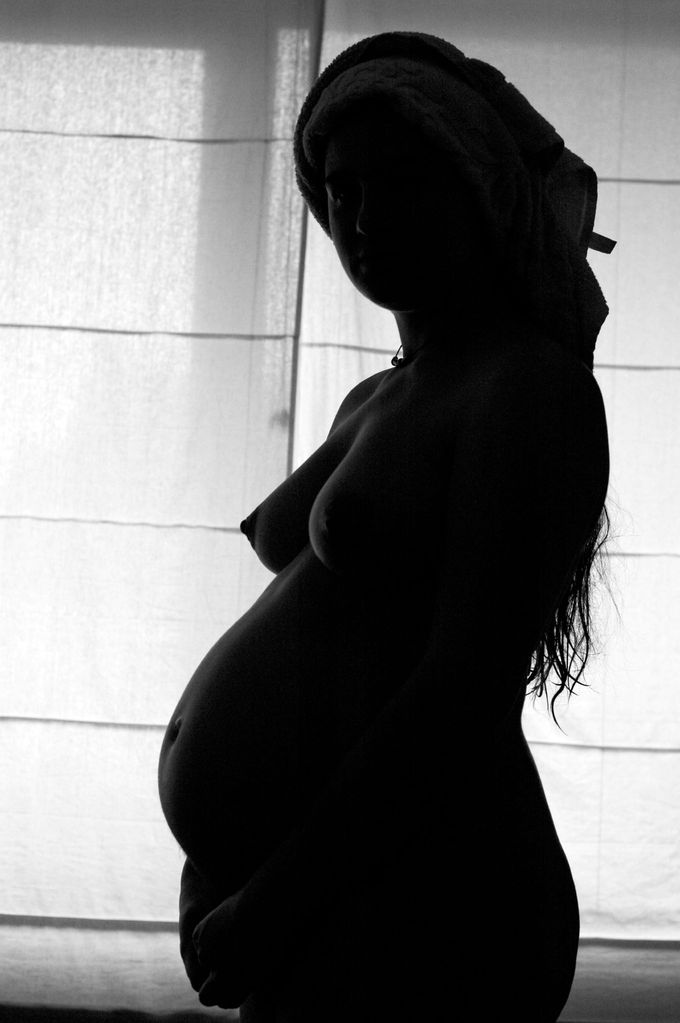
A pregnant woman: A pregnant woman’s abdomen drops due to the fetus turning in a downward position ready for birth.
The woman’s abdomen will transform in shape as the abdomen drops due to the fetus turning in a downward position ready for birth, and the woman will be able to lift her belly up and down. The woman’s navel will sometimes become convex—popping out—due to her expanding abdomen. This period of her pregnancy can be uncomfortable and cause symptoms like weak bladder control and backache.
The body’s posture changes as the pregnancy progresses. The pelvis tilts and the back arches to help keep balance. Poor posture occurs naturally from the stretching of the woman’s abdominal muscles as the fetus grows. These muscles are less able to contract and keep the lower back in proper alignment.
The pregnant woman has a different gait. The step lengthens as the pregnancy progresses due to weight gain and changes in posture. In addition, the increased body weight of pregnancy, fluid retention, and weight gain lowers the arches of the foot, further adding to the foot’s length and width.
The influences of increased hormones such as estrogen and relaxin initiate the remodeling of soft tissues, cartilage, and ligaments. Certain skeletal joints (e.g., the pubic symphysis and sacroiliac) widen or have increased laxity.
Metabolic Changes
Protein and carbohydrate metabolisms are affected during pregnancy and maternal insulin resistance can lead to gestational diabetes.
Learning Objectives
Analyze the metabolic factors involved in gestational diabetes
Key Takeaways
Key Points
- During pregnancy, the metabolism shifts to provide the growing fetus with more nutrients as well as to ensure the development of the uterine lining and breast glandular tissue.
- Hormonal changes during pregnancy increase nutrient requirements and fat deposition.
- Insulin resistance can develop and lead to gestational diabetes.
Key Terms
- cortisol: A steroid hormone (also called hydrocortisone) produced by the adrenal cortex, that regulates the metabolism of carbohydrates and maintains blood pressure.
- gestational diabetes: Also called gestational diabetes mellitus (GDM), it is a condition in which women without previously diagnosed diabetes exhibit high blood glucose levels during pregnancy (especially during the third trimester).
- lactogen: Human placental lactogen (HPL), also called human chorionic somatomammotropin (HCS), is a polypeptide placental hormone. Its structure and function is similar to that of human growth hormone. It modifies the metabolic state of the mother during pregnancy to facilitate the energy supply of the fetus.
Nutrient Metabolism
During pregnancy, both protein metabolism and carbohydrate metabolism are affected. One kilogram of extra protein is deposited, with half going to the fetus and placenta, and another half going to uterine contractile proteins, breast glandular tissue, plasma protein, and hemoglobin.
An increase in nutrients is required for fetal growth and fat deposition. Changes are caused by steroid hormones, lactogen, and cortisol. Increased liver metabolism is also seen, with increased gluconeogenesis that leads to increased maternal glucose levels. Maternal insulin resistance can lead to gestational diabetes.
Gestational Diabetes
Gestational diabetes (or gestational diabetes mellitus, GDM) is a condition in which women without previously diagnosed diabetes exhibit high blood glucose levels during pregnancy (especially during the third trimester). There is some question whether the condition is natural during pregnancy.
Maternal insulin resistance can lead to gestational diabetes. This type of diabetes is caused when the insulin receptors do not function properly. This is likely due to pregnancy-related factors, such as the presence of human placental lactogen that interferes with susceptible insulin receptors. This in turn causes inappropriately elevated blood sugar levels.
Gestational diabetes generally has few symptoms and it is most commonly diagnosed by screening during pregnancy. Diagnostic tests detect inappropriately high levels of glucose in blood samples. Gestational diabetes affects 3–10% of pregnancies, depending on the population studied, so it may be a natural phenomenon.

Effect of insulin on glucose uptake and metabolism: Insulin binds to its receptor (1) on the cell membrane which in turn starts many protein activation cascades (2). These include: the translocation of the glut-4 transporter to the plasma membrane and an influx of glucose (3), glycogen synthesis (4), glycolysis (5) and fatty acid synthesis (6).
Babies born to mothers with untreated gestational diabetes are typically at an increased risk of problems, such as being large for gestational age (which may lead to delivery complications), low blood sugar, and jaundice. If untreated, it can also cause seizures or still birth.
Gestational diabetes is a treatable condition and women who have adequate control of glucose levels can effectively decrease these risks.
Physiological Changes
Maternal physiological changes in pregnancy are entirely normal and serve as adaptations to better accommodate embryonic/fetal development.
Learning Objectives
Describe the physiological changes a woman undergoes during pregnancy
Key Takeaways
Key Points
- Women undergo several changes during pregnancy, including cardiovascular, hematologic, metabolic, renal, and respiratory changes that provide adequate nutrition and gas exchange for the developing fetus.
- Progesterone and estrogen levels rise continually through pregnancy, together with blood sugar, breathing rate, and cardiac output.
- The body’s posture changes during pregnancy to accommodate the growing fetus and the mother will experience weight gain.
- Breasts grow and change in preparation for lactation once the infant is born. Once lactation begins, the woman’s breasts swell significantly and can feel achy, lumpy, and heavy (engorgement). This is relieved by nursing the infant.
- Plasma and blood volume increase over the course of the pregnancy and lead to changes in heart rate and blood pressure. Women may also have a higher risk of blood clots, especially in the weeks following labor.
Key Terms
- human placental lactogen: Also called human chorionic somatomammotropin, this is a polypeptide placental hormone. Its structure and function is similar to that of human growth hormone. It modifies the metabolic state of the mother during pregnancy to facilitate the energy supply of the fetus.
- human chorionic gonadotropin: A peptide hormone produced during pregnancy that prevents the breakdown of the corpus luteum and maintains progesterone production.
- progesterone: A steroid hormone, secreted by the ovaries, whose function is to prepare the uterus for the implantation of a fertilized ovum and to maintain pregnancy.
Maternal physiological changes in pregnancy are the normal adaptations that a woman undergoes during pregnancy to better accommodate the embryo or fetus, and include cardiovascular, hematologic, metabolic, renal, and respiratory changes. The female body must change its physiological and homeostatic mechanisms in pregnancy to ensure proper fetal development. Increases in blood sugar, breathing, and cardiac output are all required.
Hormonal Changes
Pregnant women experience adjustments in their endocrine system. Levels of progesterone and estrogens rise continuously throughout pregnancy to suppress the hypothalamic axis and, subsequently, the menstrual cycle.
Estrogen produced by the placenta is associated with fetal well being. Women also experience an increase in human chorionic gonadotropin (β-hCG), which is produced by the placenta and maintains progesterone production by the corpus luteum.
The increase in progesterone production primarily functions to relax smooth muscles. Prolactin levels increase due to maternal pituitary gland enlargement that mediate a change in the structure of the mammary gland from ductal to lobular-alveolar.
Parathyroid hormone increases and leads to increased calcium uptake in the gut and reabsorption by the kidney. Adrenal hormones such as cortisol and aldosterone also increase.
Human placental lactogen (HPL) is produced by the placenta, stimulating lipolysis and fatty acid metabolism by the woman and conserving blood glucose for use by the fetus. It can also decrease maternal tissue sensitivity to insulin and result in gestational diabetes.
Weight Changes
One of the most noticeable alterations in pregnancy is the gain in weight. The enlarging uterus, the growing fetus, the placenta and liquor amnii, and the acquisition of fat and water retention, all contribute to weight gain.
The weight gain varies and can be anywhere from five pounds (2.3 kg) to over 100 pounds (45 kg). In the U.S., the doctor-recommended weight gain range is 25 pounds (11 kg) to 35 pounds (16 kg), less if the woman is overweight, more (up to 40 pounds 18 kg) if the woman is underweight.
Pregnancy: During pregnancy, a woman gains weight and her breasts enlarge.
A woman’s breasts grow during pregnancy, usually one to two cup sizes, but possibly larger. A woman who wore a C cup bra prior to her pregnancy may need to buy an F cup or larger bra while nursing. A women’s torso also grows and her bra band size may increase one or two sizes.
Once the baby is born (about 50 to 73 hours after birth), the mother will experience her breasts filling with milk, at which point changes in the breast happen very quickly. Once lactation begins, the woman’s breasts swell significantly and can feel achy, lumpy, and heavy (engorgement). Her breasts may increase again in size. Individual breast size can vary daily or for longer periods depending on how much the infant nurses from each breast.
Circulatory Changes
Plasma and blood volume slowly increase by 40–50% over the course of the pregnancy (due to increased aldosterone) to accommodate the changes, resulting in an increase in heart rate (15 beats/min more than usual), stroke volume, and cardiac output. Cardiac output increases by about 50%, primarily during the first trimester.
The systemic vascular resistance also drops due to the smooth muscle relaxation and overall vasodilation caused by elevated progesterone, leading to a fall in blood pressure. Diastolic blood pressure consequently decreases between 12–26 weeks, and increases again to pre-pregnancy levels by 36 weeks.
Edema (swelling) of the feet is common during pregnancy, partly because the enlarging uterus compresses veins and lymphatic drainage from the legs.
A pregnant woman will also become hypercoagulable, leading to increased risk for developing blood clots and embolisms due to increased liver production of coagulation factors. Women are at highest risk for developing clots (thrombi) during the weeks following labor.
Clots usually develop in the left leg or the left iliac venous system because the left iliac vein is crossed by the right iliac artery. The increased flow in the right iliac artery after birth compresses the left iliac vein leading to an increased risk for thrombosis (clotting) that is exacerbated by a lack of ambulation (walking) following delivery. Both underlying thrombophilia and caesarean section can further increase these risks.
Exercise and Pregnancy
In the absence of complications, pregnant women should continue aerobic and strength training exercise for the duration of gestation.
Learning Objectives
Evaluate the types of exercise appropriate for pregnant women
Key Takeaways
Key Points
- Moderate aerobic exercise and strength training improve the health of pregnant women while having no adverse consequences on the developing fetus.
- A variety of exercise activities are appropriate, with the exception of those with a high risk for abdominal trauma, such as horseback riding, skiing, soccer, or hockey.
- Contraindications of exercise include: vaginal bleeding, dyspnea before exertion, dizziness, headache, chest pain, muscle weakness, preterm labor, decreased fetal movement, amniotic fluid leakage, and calf pain or swelling (to rule out thrombophlebitis).
Key Terms
- contraindication: A factor or symptom that makes the prescribed treatment inadvisable.
- aerobic exercise: Physical exercise of low to high intensity that depends primarily on the aerobic energy-generating process.
- strength-conditioning: The use of resistance to muscular contraction to build the strength, anaerobic endurance, and size of skeletal muscles. There are many different methods of strength training, the most common being the use of gravity or elastic/hydraulic forces to oppose muscle contraction.
- thrombophlebitis: Phlebitis (vein inflammation) related to a thrombus (blood clot).

Exercising while pregnant: A strong, healthy woman will generally have a good pregnancy outcome. Physicians recommend moderate exercise during pregnancy, including strength-training.
Regular aerobic exercise during pregnancy appears to improve (or maintain) physical fitness. Although an upper level of safe exercise intensity has not been established, women who were regular exercisers before pregnancy and who have uncomplicated, healthy pregnancies should be able to engage in high-intensity exercise programs (e.g., jogging and aerobics) for less than 45 minutes with no adverse effects. They just need to be mindful of the possibility that they may need to increase their energy intake, and are careful to not become overheated.
In the absence of either medical or obstetric complications, doctors advise an accumulation of 30 minutes a day of exercise on most if not all days of the week. The Clinical Practice Obstetrics Committee of Canada recommends that “all women without contraindications should be encouraged to participate in aerobic and strength-conditioning exercises as part of a healthy lifestyle during their pregnancy.”
In general, participation in a wide range of recreational activities appears to be safe. Pregnant women just need to avoid those with a high risk of falling such as horseback riding or skiing, or those that carry a risk of abdominal trauma, such as soccer or hockey.
In the past, the main concerns of exercise in pregnancy were focused on the fetus and any potential maternal benefit was thought to be offset by potential risks to the fetus. However, more recent information suggests that in the uncomplicated pregnancy, fetal injuries are highly unlikely.
Contraindications for exercise include, vaginal bleeding, dyspnea before exertion, dizziness, headache, chest pain, muscle weakness, preterm labor, decreased fetal movement, amniotic fluid leakage, and calf pain or swelling (to rule out thrombophlebitis).
