Human Anatomy Reading – Anatomy of Skin
Learning Objectives
Test questions are written using concepts listed in the Learning Objectives.
- Describe the structure of the skin and the tissue types that comprise it. Distinguish between the epidermis, dermis, and hypodermis.
- Describe the characteristics of the stratum basale and stratum corneum in the epidermis. Compare and contrast the characteristics and functions of the keratinocytes in each layer.
- Describe the functions and locations of melanocytes, Langerhans cells, and Merkel cells.
- Compare and contrast the layers of the skin that are involved in first, second and third degree burns as well as the basic clinical consequences of each type of burn.
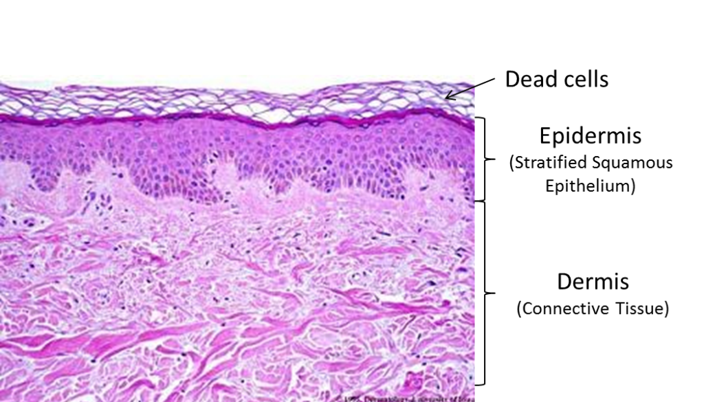
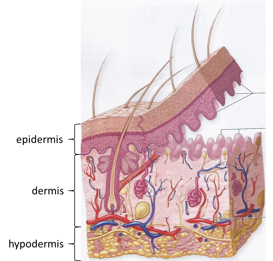
The skin is the organ of the body with which we are most familiar, and the one we most take for granted. It has many crucial functions. It forms a barrier to abrasion, infection, UV rays, and dehydration; it is a sensory organ and an excretory organ; it helps regulate body temperature and is necessary for the production of vitamin D. Structurally, skin has two layers: the epidermis, keratinized stratified squamous epithelium, and the dermis, a connective tissue network of collagen and elastic fibers containing blood vessels and nerve endings. The structure of the skin is highly specialized by region. In areas with increased friction, it is thicker and tightly fastened to underlying tissues. In areas where it is involved in grasping, the skin has prominent ridges (look at your fingertips!).
KNOWLEDGE CHECK
Structure of the Skin
The skin is composed of two tissue layers, the outer epidermis and the inner dermis. The average thickness of the two layers is about 1/8 inch, but the actual thickness varies a great deal, based on the functional needs of different areas of skin. The tissue layer deep to the dermis is called hypodermis. It is not considered to be part of the skin ‘proper.’ It is made primarily of adipose tissue and it varies greatly in thickness regionally (and between individuals).
KNOWLEDGE CHECK
Innervation of the Skin
As stated previously, innervation to the skin travels to and from the spinal cord via the spinal nerves and their branches, the dorsal and ventral rami. Innervation of the skin is segmental. Each spinal cord level receives sensory information from one strip of skin, a dermatome.
Nerves in the skin are cutaneous nerves and contain sensory and sympathetic fibers. Sensory fibers convey all sensation from the skin to the central nervous system (pain, touch, and temperature). Sympathetic fibers innervate sweat glands, arrector pili muscles, and the blood vessels of the dermis. Almost all innervation to the skin is in the dermis. With the exception of a few free nerve endings, the nerves do not reach the epidermis.
KNOWLEDGE CHECK
Burns
Burns are among the most traumatic of injuries. Besides causing tissue damage, burns breach the barrier functions of the skin, exposing the body to microorganisms and allowing significant fluid loss from the body, which can disrupt blood volume and organ function. Major burns cause homeostatic imbalances in every system of the body, a testament to the importance of skin function to the body.
One scheme for classifying burns is based on how many layers of tissue are injured:
- In first-degree burns, the epidermis is damaged, but not destroyed. First-degree burns are red and painful, but they are not serious. There may be swelling, redness and pain. The injured cells peel off and the skin will heal, without scarring, in about two weeks.
- Second-degree burns destroy the epidermis and also cause some cell destruction in the dermis. Blisters appear and the surface is usually wet or moist due to plasma loss. There may be greater pain with second-degree burns than with third-degree burns (the most severe) because nerve endings in second-degree burns are still intact, but damaged. New skin can regenerate from the remaining dermis, but there may be scarring. All second-degree burns should receive medical attention.
- Third-degree burns destroy the epidermis and dermis, and can extend into the underlying hypodermis. Because nerve endings in the dermal layer of the skin are destroyed in third-degree burns, these burns may not be as painful as less severe burns. The burned area will be relatively dry, due to the loss of the blood vessels of the dermis, and often has a white or charred appearance. Since the dermis is destroyed, the skin cannot regenerate. Surgery and skin grafts are always necessary.
KNOWLEDGE CHECK
